Single photon pulses were measured at different wavelength setting of
the monochromator from the UV (200 nm) to the near infra-red (![]() m).
Sample spectra are shown in Fig. 2 (click here). The mean charge output <Q> and the FWHM
m).
Sample spectra are shown in Fig. 2 (click here). The mean charge output <Q> and the FWHM
![]() provide a determination of the detector responsivity
provide a determination of the detector responsivity ![]() and
measured spectral resolution
and
measured spectral resolution ![]() for a known photon
wavelength. A typical responsivity of
for a known photon
wavelength. A typical responsivity of ![]() electrons/eV,
equivalent to a mean number of tunnels for each charge carrier of
electrons/eV,
equivalent to a mean number of tunnels for each charge carrier of ![]() , was derived. The peak at the end of the charge range in the spectrum
of Fig. 2 (click here)a is due to the pulsar used to determine the contribution to the
measured resolution from the electronics
, was derived. The peak at the end of the charge range in the spectrum
of Fig. 2 (click here)a is due to the pulsar used to determine the contribution to the
measured resolution from the electronics ![]() . At some wavelengths the
second and third order from the monochromator are also apparent rather
nicely illustrating the detectors intrinsic spectroscopic capability. The
short and long wavelength limits of 200 nm and
. At some wavelengths the
second and third order from the monochromator are also apparent rather
nicely illustrating the detectors intrinsic spectroscopic capability. The
short and long wavelength limits of 200 nm and ![]() are due to the fibre
optic cut-off.
are due to the fibre
optic cut-off.
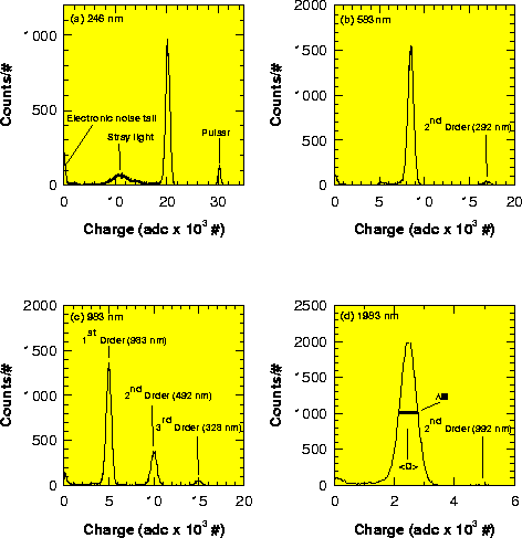
Figure 2: The charge spectra obtained from the junction when illuminated with
a) 246 nm , b) 583 nm, c) 983 nm and d)
1983 nm photons from a
monochromatic light source. The charge is in units of ADC channel (#) as
recorded by the signal pulse height analyser ![]() electrons)
electrons)
![]()
Figure 3: The measured resolution ![]()
![]() and the electronics corrected
resolution
and the electronics corrected
resolution ![]()
![]() as a function of wavelength together with the best
linear fits. The theoretical variation with photon wavelength of the Fano
plus tunnel noise limited resolution
as a function of wavelength together with the best
linear fits. The theoretical variation with photon wavelength of the Fano
plus tunnel noise limited resolution ![]() )
based on Eq. (2) is also
shown. The inset shows the current detectors signal to noise ratio (S/N) as
a function of the near infrared photon wavelength in
)
based on Eq. (2) is also
shown. The inset shows the current detectors signal to noise ratio (S/N) as
a function of the near infrared photon wavelength in ![]() m
m
Figure 3 (click here) illustrates the measured resolution ![]() of the device as a
function of wavelength together with the resolution
of the device as a
function of wavelength together with the resolution ![]() after correction
for the electronic noise
after correction
for the electronic noise ![]() ), as determined from the constant charge
input pulse, such that
), as determined from the constant charge
input pulse, such that ![]() . The limiting resolution of the
device
. The limiting resolution of the
device ![]() ) is also shown in Fig. 3 (click here),
based on Eq. (2) with
) is also shown in Fig. 3 (click here),
based on Eq. (2) with ![]() and
a value of
and
a value of ![]() , corresponding to that of bulk tantalum. The agreement
between
, corresponding to that of bulk tantalum. The agreement
between ![]() and
and ![]() is very good indeed, indicating that the detector
resolution at these wavelengths is totally dominated by the tunnel noise
with little contribution from spatial variations to the responsivity
is very good indeed, indicating that the detector
resolution at these wavelengths is totally dominated by the tunnel noise
with little contribution from spatial variations to the responsivity
![]() )
as observed at X-ray wavelengths (Verhoeve et al. 1996, 1997). In
addition the signal to noise ratio is also shown in Fig. 3 (click here) (inset).This
allows an estimate of the current long wavelength response beyond that
actually measured of
)
as observed at X-ray wavelengths (Verhoeve et al. 1996, 1997). In
addition the signal to noise ratio is also shown in Fig. 3 (click here) (inset).This
allows an estimate of the current long wavelength response beyond that
actually measured of ![]() m. Assuming a five sigma detection criterion
above the noise the detector is currently limited by the electronic noise
to wavelengths up to
m. Assuming a five sigma detection criterion
above the noise the detector is currently limited by the electronic noise
to wavelengths up to ![]() m. Note this maximum wavelength
m. Note this maximum wavelength
![]() can be simply written as
can be simply written as ![]() ]. Further reduction in
the noise of the room temperature electronics would of course extend even
further this value into the infrared.
]. Further reduction in
the noise of the room temperature electronics would of course extend even
further this value into the infrared.
The linearity of the detector with photon energy is best measured using the multiple orders from the grating. This removes effects such as response changes with drifting temperature or possible calibration errors since the output from the monochromator over several grating orders are exact multiples of the same wavelength. Figure 4 (click here) shows the charge spectrum from the device when illuminated with optical light via the grating monochromator, with such a grating response covering four orders from 1183 - 296 nm. Not only are the various orders well resolved but the charge output as a function of wavelength can be precisely determined. The linearity is very high with a maximum deviation from linearity with photon energy of below 0.6%.
![]()
Figure 4: The complete charge spectrum when the monochromator was set to
select photons with a wavelength of 1183 nm. The grating orders 1 to 4 are
all clearly resolved. Some distortion in the charge distribution at low and
possibly higher charge levels can be discerned in each of these peaks and
may be due to pile-up of events due to the signal process time of the
electronics. Stray light and the incomplete conversion of the photons energy
to charge carriers, due to possible substrate losses, have been ruled out as
a major cause of such low intensity level tails in these spectra